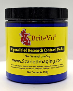
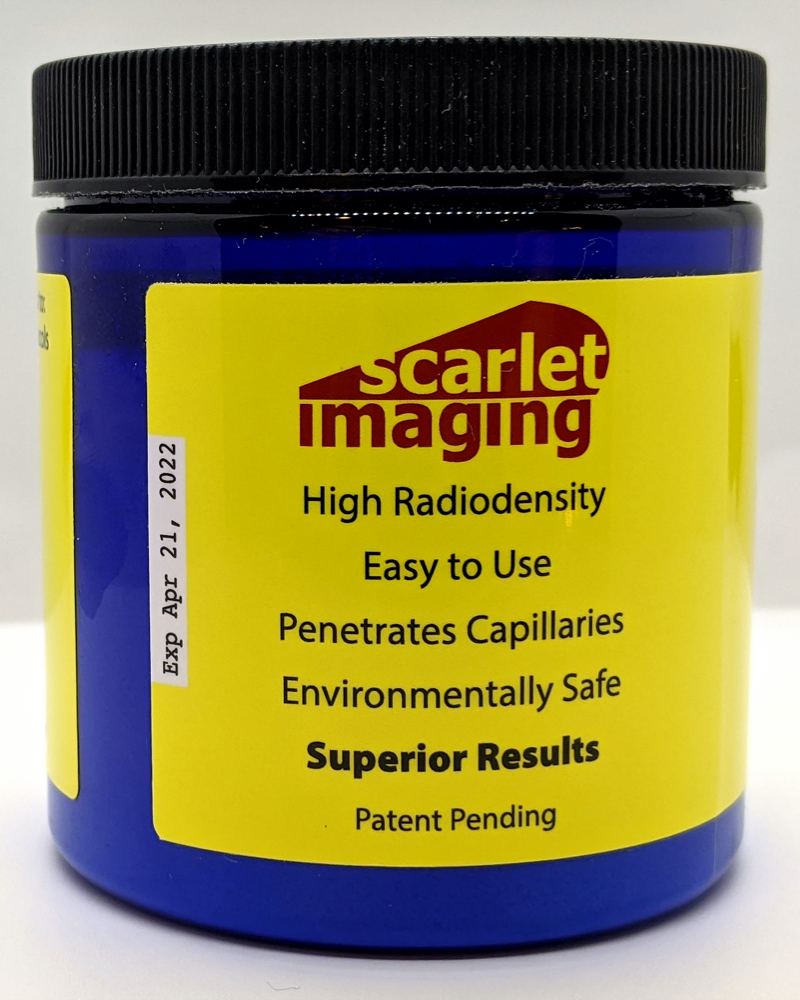
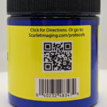
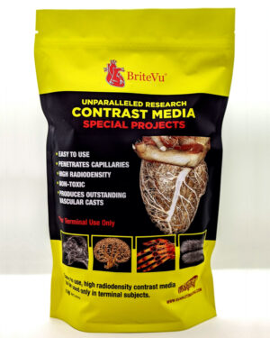
BriteVu 170g
$154.99
Buy 4 jars and get 10% off product price
($139.49* per 170g jar)
Buy 5 or more jars and get 20% off product price!
($123.99* per 170g jar)
Price includes FedEx 3-5 day shipping for ALL DOMESTIC orders. For EXPEDITED or INTERNATIONAL shipping, please Contact us for quotes.
***PLEASE NOTE: Customer is responsible for customs duties and taxes for international shipments.
Please allow 2-4 business days to process order prior to shipping. Contact us for other shipping arrangements (especially rush orders or using your own carrier).
Description
BriteVu 170g Basics:
BriteVu is a high radiodensity contrast media to be used only in terminal subjects. Each 8oz jar contains 170 g of powdered material. BriteVu (1 part in grams) is mixed with distilled water or other select water soluble solvents (3-4.5 parts in ml at full strength). When properly mixed, each jar can produce approximately 510-765 ml of smooth white liquid contrast material. This is enough material to perfuse 10-15 adult mice and 5-9 adult rats (< 300 g).
The contents of each jar are mixed per the directions below and perfused into the test subject. Perfuse the product into the system being evaluated. For example, for blood vessel evaluation perfuse BriteVu intravascularly. As a note, protocols can vary significantly based on intended results. The protocol below provides the most common guidelines for preparation.
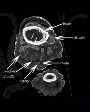
The perfused subject is later scanned using radiographs, computed tomography (CT), magnetic resonance imaging (MRI) or ultrasound depending on the study.
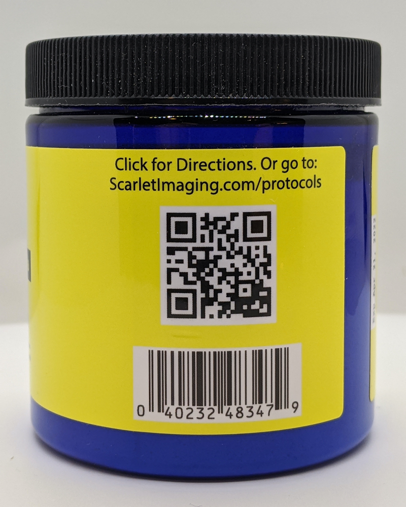
Item #: 040232483479
BriteVu Compatibility:
-Computed Tomography (CT)
-Magnetic Resonance Imaging (MRI)
-Ultrasound
-Photography and Videography
Preparation Directions:
For Best Results Mix 1 Part BriteVu (in grams) to 3.0-4.5 Parts Distilled Water (in cc):
(As an example, mix 25 grams of BriteVu in 75-113 cc of water)
- Heat the appropriate amount of distilled water in a glass beaker with mixing rod to at least 40-45 deg C on a stirring hot plate.
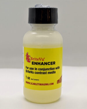
- If using BriteVu Enhancer, add to the warm water and mix for at least 1 minute.
- Add the appropriate amount of BriteVu into the heated water bath with mixing rod stirring such that all powder goes into solution.
- Heat solution to 70-80 deg C for at least 10 minutes.
- Immediately perfuse the entire solution at 45-65 deg C into the deceased subject. If using BriteVu Enhancer, perfusions can be performed at 35-55 deg C preserving tissue histology.
- Cool the perfused subject (ice water bath, refrigerator, etc) until BriteVu mixture solidifies and then perform imaging studies.
- Alternatively, the cooled subject and/or its tissues can be stored in fixative for later scanning.
- Protocols can vary based on intended results. Go to Protocols for more details.
BriteVu Advantages:
-Completely non-toxic.
-Easy to use.
-Penetrates to the capillary level.
-Produces outstanding casts compatible with CT, MRI, ultrasound, photgraphy and videography.
Comparison of Commercially Available Terminal Vascular Contrast Agents
Shipping Information:
***PLEASE NOTE: Customer is responsible for customs, duties and taxes for international shipments.
Please allow 2-4 business days to process order prior to shipping. Contact us for other shipping arrangements (especially rush orders or using your own carrier).
Additional information
| Weight | 200 g |
|---|---|
| Dimensions | 2.75 × 2.75 × 3 in |
3 reviews for BriteVu 170g
You must be logged in to post a review.

scarlet –
BriteVu is one of the most exciting and innovative things to happen to the field of anatomy in a long time. Everything about BriteVu is an upgrade from the current products out there for vascular imaging, and the staff at Scarlet Imaging are both wonderful to work with and willing bend over backwards to help facilitate the research needs of their clients.
Emma R. Schachner, PhD
Laboratory for Equine and Comparative Orthopedic Research, Department of Veterinary Clinical Sciences, School of Veterinary Medicine, Louisiana State University
scarlet –
BriteVu provides remarkable vascular contrast in X-ray images, particularly across small-vessel lumina. It’s an easy-to-use visualization tool with predictably clear results.
Paul M. Gignac, PhD
Department of Anatomy & Cell Biology, Oklahoma State University Center for Health Sciences
scarlet –
An Aneurism is a localized, blood-filled balloon-like bulge in the wall of a blood vessel. When the size of an aneurysm increases, there is a significant risk of rupture, resulting in severe hemorrhage, and other complications, even death. The exact cause of an aneurysm is unclear. Incidence rate of aneurysm in infant could be up to 3.6%, and often associated with other congenital heart defects. In adults, aneurysm formation is probably the result of multiple factors affecting the arterial segment and its local environment. To understand the morphogenesis of cardiac anomalies, specifically the homeostasis of the blood flow resulting from an aneurysm, part of our research includes the injection of BriteVu high radiodensity contrast media to understand the 3-dimension cardiovascular system resulted from the disease via micro-computed tomography (µ-CT). Since we are utilizing chick embryo to study the early cardiovascular development, the BriteVu contrast media is readily soluble in water and fairly stable in the colloidal liquid when maintained in a warm incubator for a long period of time. This is essential for easy injection to the tiny vascular system since the whole embryo could be merely few millimeter of blood volume. Most of all, the media provides fine details of the heart structure and its vasculature from µ-CT scan.
Norman Hu, MS
Department of Pediatrics, University of Utah