What is BriteVu
BriteVu is a radiodense contrast agent for use in terminal research subjects and cadavers.
How to use BriteVu
BriteVu comes as a powder and is mixed with water, phenol and/or other select water soluble solvents to form a solution that is perfused and forms a solid gel once cooled. The high radiodensity of BriteVu allows one to see the perfused system using computed tomography (CT), radiographs (x-rays) and with some magnetic resonance imaging (MRI) scans. Subjects can be imaged whole and/or fixed organs/tissues can be scanned individually.
For specific details on how to use BriteVu with various subjects, see the BriteVu Perfusion protocol. If you have other subjects or ideas not covered in the BriteVu Perfusion Protocol, please Contact us. We would love to hear about your project!
BriteVu – Helping researchers achieve better answers that change the world.
What can it be used for?
BriteVu is primarily used as an intravascular agent to cast the cardiovascular system down to the capillary level to be visualized using various imaging modalities (CAT scans, x-rays, etc). However, BriteVu can be injected into any cavity including the respiratory and gastrointestinal systems, cerebrospinal fluid, body compartments and more. Any animal can conceivably be perfused with BriteVu. BriteVu can be also be used to fill compartments of mechanical systems and other non-biologic subjects.
Specific areas where BriteVu can be used for teaching include the following (and more):
- Anatomic discovery (different species and anatomic locations not previously defined)
- Artwork
- Delineating vascular anatomy of whole organisms and individual organs and tissues
- Developing teaching models
- -3-D computer models
- -3-D digitally printed models
- -3-D hologram models
- Flat images for publications
- Visual aids for all levels of education
- Understanding physiology
BriteVu advantages
Comparison of Commercially Available Terminal Vascular Contrast Agents
Usage Examples:
Birds: Back
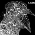
Pigeon Head High Radiodensity Contrast Perfusion Using BriteVu
The vasculature throughout the head and beak (rostral) can easily be seen. Such studies allow anatomists to clearly trace the vascular pathways in 3D and show blood supply to various tissues in complex regions such as the head. The pigeon was CT scanned on the Siemens Inveon Micro CT at 100 µm.
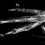
Pigeon Foot High Radiodensity Contrast Perfusion Using BriteVu
It is difficult to get vascular anatomy of small extremities such as that with pigeon toes. However, BriteVu readily penetrates even the most distal extremities creating beautiful studies. Digits 2-4 are visible in this image. CT scanned on the Siemens Inveon Micro CT at 100 µm.
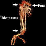
Grey Parrot Leg High Radiodensity Contrast Perfusion Using BriteVu
This particular study shows the complex vasculature present particularly at the caudal angle of the stifle in the grey parrot. This correlates with the thick muscle mass present in this location. The leg was scanned on the Siemens Inveon Micro CT and digitally segmented out of a 100 µm slice CT scan.
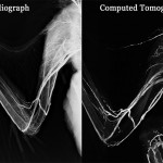
Golden Eagle Wing High Radiodensity Angiogram Using BriteVu
This image on the left comes from a standard digital radiograph of the wing and the one on the right from a 500 µm CT scan. Immediately after perfusion, the bird was radiographed and then transferred to a CT center for additional imaging. These studies give clinicians insight as to how vasculature may appear (with live animal contrast agents) on flat 2D radiographs compared with 3D CT. This information is useful to better understand basic anatomic location of vessels and also spatial positioning of the vasculature.
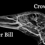
Pekin Duck Head High Radiodensity Contrast CT Scan Using BriteVu
Head anatomy is incredibly complex in many animals. Because of the highly variable beak apparatus across the avian species, the vascular anatomy is also quite unique. Compare the duck, pigeon and grey parrot head anatomy featured in this section- there are tremendous differences in vascular anatomy. CT scanned at 100 µm on the Siemens Inveon Micro CT.
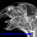
Grey Parrot Head Contrast CT Scan Using High Radiodensity Agent (BriteVu)
Notice the ‘kink’ in the carotid artery supplying blood to the head. This kink or slack is found in many bird species that are capable of turning their head around. Without this extra vascular length, the bird would likely pass out if turning its head too far. This is a great example of the vascular adaptation to the bird’s form and function. This bird was CT scanned (whole body) at 100 µm on the Siemens Inveon Micro CT.
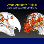
3D Model of Parrot Head Perfused with BriteVu
Using a variety of digital subtraction tools, the blood vessels of the grey parrot head can be viewed with and without their boney coverings. The image on the right shows a sagittal section through the beak and skull with vessels readily visible. These teaching models are invaluable when demonstrating the location of various vessels (and their associated tissues) within bone or other coverings. These models also help guide clinicians when determining where to collect fluid samples or make surgical incisions.
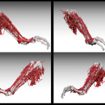
3D Model of Grey Parrot Leg Perfused with BriteVu
Courtesy of Scott Birch, The Center for Educational Technologies, Texas A&M College of Veterinary Medicine & Biomedical Sciences. Scott took the data sets of a BriteVu perfused grey parrot to create 3-D models of the vasculature and bone. These were his first pass images!
Humans: Back
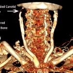
Human neck perfused with BriteVu
Human neck region perfused with BriteVu.
This cadaver was perfused with BriteVu Special Projects to reveal the blood vessels throughout the body (with the help of a CAT scan!). There are several interesting abnormal findings here: a broken right collar bone, occluded right carotid artery (likely from atherosclerosis) and a set of surgical steel rods in the back of the spine. Perfusion and CT scan courtesy of Dr Bruce Wainman, Department of Education, McMaster University and Touch of Life.
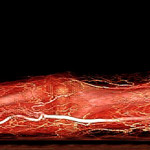
Human arm perfused with BriteVu contrast agent
The shoulder to the fingers plus the entire associated vasculature are visible. The radial artery is seen coursing along forearm (bright vessel) and over the dorsal base of the thumb. The arm was CT scanned at 2 mm. Courtesy of Dr. Bruce Wainman, Education Program in Anatomy, McMaster University.
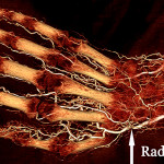
Human hand perfused with BriteVu contrast agent
The distal radius and ulna bones plus the entire hand and vasculature are visible. Again, the radial artery is highlighted. The hand was CT scanned at 2 mm. Courtesy of Dr. Bruce Wainman, Education Program in Anatomy, McMaster University.
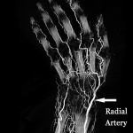
BriteVu Perfused Human Wrist
In line with the other images of the hand, the radial artery is highlighted. Understanding the 3D orientation of the hand and digital vasculature becomes critical in the case of injuries or surgery in this area. Clinicians need this information when making clinical-surgical decisions. Contrast arteriograms created in part by BriteVu can make incredible 3D digital atlases that can be used in the classroom, research and in clinical practice. The hand was CT scanned at 2 mm. Courtesy of Dr. Bruce Wainman, Education Program in Anatomy, McMaster University.
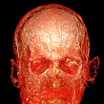
BriteVu Perfused Human Head
This anterior (rostral) posterior view (front to back) shows the vasculature of the readily recognizable eye sockets, forehead and ears. The lips can be seen at the bottom of the image. The head was CT scanned at 1 mm slice thickness. Courtesy of Dr. Bruce Wainman, Education Program in Anatomy, McMaster University.
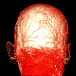
BriteVu Perfused Human Head
This posterior anterior (rostral) view (back to front) shows the vasculature of the readily recognizable ears as well as the top of the head and back of the neck. The head was CT scanned at 1 mm slice thickness. Courtesy of Dr. Bruce Wainman, Education Program in Anatomy, McMaster University.
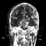
BriteVu Perfused Human Head- Coronal Section
This slightly rotated 2.5 cm (1 inch) coronal section through the skull at the level of the ears shows the vasculature of the brain (top), ear canals and a portion of the upper spine. 3D image manipulation technology allows users to study very specific portions of the subject’s body. The arteriovenogram using BriteVu allows users to see the vasculature with incredible detail. The head was CT scanned at 1 mm slice thickness. Courtesy of Dr. Bruce Wainman, Education Program in Anatomy, McMaster University.
Non-Human Mammals: Back
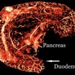
Mouse Duodenum and Pancreas Perfused with BriteVu
The anatomic arrangement of the pancreas and its vascular relationship to surrounding organs is important to studying pancreatic diseases, especially diabetes mellitus, and understanding its unique anatomy. With the addition of BriteVu contrast agent, researchers and teachers alike can easily visualize the pancreas vascularity and its direct blood supply relationship with the intestines (duodenum).
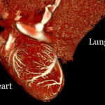
BriteVu Perfused Contrast Scan of a Rat Heart and Lung
With the newer 3-D imaging technology, teachers can take data sets from studies such as this BriteVu perfused rat heart and lungs and create amazing classroom learning tools. Even with this small rat heart and lungs, details can be seen down to the capillary level. The bright contrast between the BriteVu filled vessels and the heart and lung tissue make digital segmenting easy. From here, 3-D printed models and imaging tools can be used to teach students about blood flow and vascular relationships between organs.
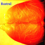
BriteVu Perfused Contrast Agent in the Rat Brain
Stewart Yeoh from the Head Injury and Vessel Biomechanics Lab at the University of Utah provided this image of a BriteVu perfused rat brain. This is plain light photography of the dorsal surface of the brain. The surface vessels are readily visible and contain BriteVu high contrast agent. The vasculature inside the brain tissue is also well perfused and can be seen in the light histology slide also provided by Stewart Yeoh in the ‘For Research’, ‘Neurovascular’ gallery.
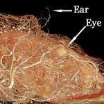
High Radiodensity Contrast Scan of a Rat Head Using BriteVu
The rat head was scanned at 35 µm resolution on the Siemens Inveon Micro CT. The nose is to the right and eye is approximately in the center of the image. Because BriteVu can be used to perfuse an entire body, complex vascular structures (such as that found in the head) can be digitally segmented and brought to life for a multitude of teaching purposes. Courtesy of Stewart Yeoh from the Head Injury and Vessel Biomechanics Lab at the University of Utah.
High Radiodensity Contrast Scan of a Rat Head Using BriteVu
Heads are commonly used as teaching models due to their complexity. The resolution and vessel size visible depends on the CT scanner resolution and slice thickness. The smaller the slice thickness the more (and smaller) vessels you will see filled with BriteVu. The head was scanned at 70 µm resolution on the Siemens Inveon Micro CT. This a lateral view with the rat’s nose to the right.
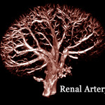
Rat Kidney High Radiodensity CT Angiogram Using BriteVu
Better known and the ‘broccoli view’! The kidney was positioned to highlight the renal artery (base or stalk of the ‘broccoli’) as it enters and supplies blood to the kidney. Scanned on the Siemens Inveon Micro CT.
Reptiles: Back
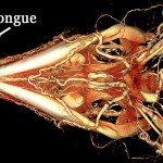
Argentine Black and White Tegu Head perfused with BriteVu
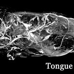
Courtesy of Dr Colleen Farmer, Department of Biology, University of Utah. This is a ventral (underside) view of the skull showing all of the blood vessels. Notice the rich blood supply around the powerful lower jaws (mandibles). The tongue can be seen coursing out of the tip of the snout (and aimed toward the top of the picture). BriteVu Rat was used in the perfusion process. This is a 100 µm CT scan using the Siemens Inveon Micro CT.
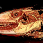
Argentine Black and White Tegu BriteVu Perfusion of Head
Courtesy of Dr Colleen Farmer, Department of Biology, University of Utah. The vasculature is seen all the way to the tip of the tongue. There are several interesting anatomic features of this animal including a radiating vascular ring inside both nares. This is a lateral view of the head. This is a 100 µm CT scan using the Siemens Inveon Micro CT.
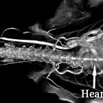
Alligator Heart and Neck High Radiodensity Contrast Perfusion with BriteVu
The alligator was scanned at 100 µm. This dorsal-ventral (top down) view shows the vessels coursing to the neck forelimbs, spinal column and caudal coelom (lower abdomen). Courtesy of Dr Colleen Farmer, Farmer Lab, University of Utah. Scanned on Siemens Inveon Micro CT.
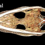
Alligator Head High Radiodensity Contrast Perfusion with BriteVu
The alligator was CT scanned at 100 µm. This is a ventral-dorsal (bottom up) view. The blood supply entering the skull (right side of picture) can be readily seen. Interestingly, the vascular supply to the alligator skull is much less obvious than that seen in other reptiles. It may be that the thick alligator skull bone obscures visualization of smaller vessels. Courtesy of Dr Colleen Farmer, Farmer Lab, University of Utah. Scanned on Siemens Inveon Micro CT.
White Throated Monitor High Radiodensity Contrast Perfusion with BriteVu
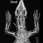
The lizard was CT scanned at 300 µm. This view and the following of the same animal offer 180 degree angle images of the vasculature. This is a ventral-dorsal (or bottom up) view.
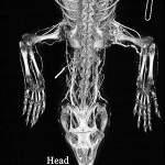
White Throated Monitor High Radiodensity Contrast Perfusion Using BriteVu
White throated monitor whole body arteriovenogram using BriteVu contrast media. The lizard was CT scanned at 300 µm. Compare with the ventral-dorsal view above. This is a dorsal-ventral (or top down) view.
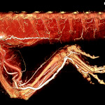
Bearded Dragon Leg and Tail Perfused with High Radiodensity Contrast Media (BriteVu)
The tail is seen coursing along the top edge of the image. The leg and it vasculature are readily apparent. The lizard was CT scanned at 100 µm on the Siemens Inveon Micro CT.

